Mounting spacers: ANTI – FLATTENING

Mounting media with microscopic spacers
Cell flattening during slide preparation isn’t solely due to hardening mounting media. The capillary pressure between the microscope slide and the cover glass also contributes to this effect. High viscosity mounting medium penetrates biological structures slower and cover glass + slide initiate capillary pressure. This causes the cell flattening.
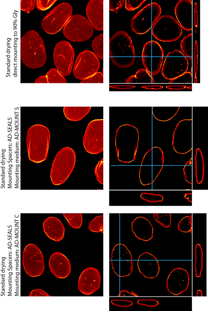
Using non-hardening mounting media AD-MOUNT and precise microscopic spacers AD-SEAL helps maintain the natural shape of biological objects. This is crucial for certain types of analysis or imaging. The sample preparation technique also prevents deformation or damage larger biological structures.
In the left image, we see pronounced cell flattening due to a common practice of cell drying (intended to remove residual water, preventing refractive index mismatch, and minimizing spherical aberration as per the Mounting procedure protocol). When using a 90% glycerol viscosity mounting medium, the flattening effect is also noticeable. However, when combining AD-MOUNT media and AD-SEAL microscopic spacer, this flattening effect is virtually eliminated.
