
Mounting media – Protect your signal, support your imaging
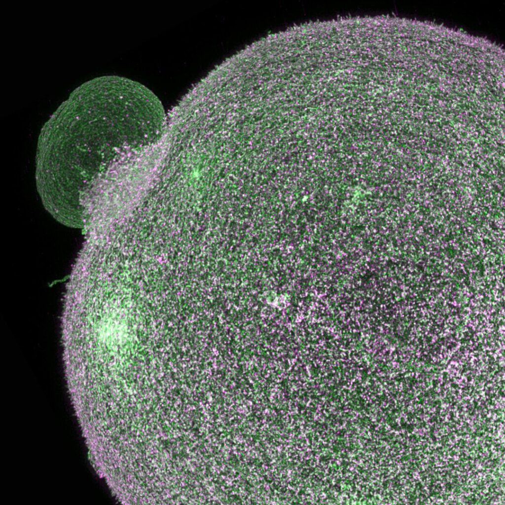
This 3D STED image shows the full structure of a mouse oocyte, labelled with two surface proteins.
Juno (green) and CD9 (magenta) highlight fine microvilli across the oocyte membrane.
Used materials: mounting medium AD-MOUNT C, mounting spacers AD-SEAL.
Biological samples distort light.
Cells and tissues are optically inhomogeneous—unlike glass or water, they bend light unevenly. When the refractive index of the sample doesn’t match its surroundings, light paths become distorted. The result: blurred images, reduced contrast, and unwanted halos that obscure fine detail.
Mounting media make images sharp and stable.
A good mounting medium penetrates deep into the sample and optically “levels out” these differences. It minimizes refractive index mismatch to improve clarity and resolution. At the same time, it protects fluorescent dyes from photobleaching and helps preserve signal intensity—so your sample stays bright and usable even after long-term storage at 4 °C.
For best results, combine with mounting spacers.
They help control sample thickness, prevent compression, and ensure consistent imaging conditions. [Learn more →]
Want to know more?
See the greatest achievement:
3D STED image of entire mouse oocyte
Learn how the sample preparation allows incredibly clear super-resolution imaging deep in the specimen.
Or download summarized bleaching assay as a PDF
Watch our short video protocol:
Mounting procedure
Your primary choices are AD-MOUNT F (non-hardening) and AD-MOUNT H (hardening) – high-quality mounting media available in both DAPI and non-DAPI variants. Both options offer excellent fluorescence stabilization, easy handling, and are fully comparable to leading international standards.
AD-MOUNT S is an advanced medium with outstanding anti-fade properties. Fluorescent signal remains extremely stable and highly resistant to photobleaching. In some cases, signal from expressed proteins may appear weaker – we recommend testing the medium beforehand.
AD-MOUNT C is designed for specialized applications. It offers a refractive index matching that of borosilicate glass, significantly reducing spherical aberration. Its mild clearing effect allows for remarkable visualization of deep structures. As with AD-MOUNT S, signal from expressed proteins may be reduced – we recommend testing.
Note: this variant is not compatible with SiR staining.

Universal Application
Refractive index: 1.45
Hardening
All fluorophores
Universal hardening mounting medium for all fluorophore types. It offers long term stabilization of structures (like phalloidin-stained actin), dyes, minimizing photo-bleaching and fluorescence fading.
Designed for long storage of samples.
Universal Application
Refractive index: 1.45
Non-hardening
All fluorophores
Universal mounting medium accommodates a wide-range of fluorophore types. It offers optimized photo-bleaching and fluorescence fading, effectively preserving an integrity and a shape of biological structures.
Designed for long storage of samples.
Superior Stability
Refractive index: 1.47
Non-hardening
Synthetic dyes
Superior Stability mounting medium ensures robust stabilization of fluorescence signal, minimizes photo bleaching in all commonly used synthetic dyes. Preserves the integrity and shape of biological structures.
Designed for long storage of samples.
Clear view
Refractive index: 1.518
Non-hardening
Synthetic dyes
Specifically formulated for high- and super-resolution microscopy. High refractive index, exceptional clearing capabilities, and fluorescence signal stabilization effect. Suitable for commonly used synthetic dyes, but not suitable for expressions and SiR.

For non-hardening mounting media, the use of mounting spacers is crucial to maintaining the natural shape and volume of biological samples. Spacers create a controlled gap between the cover slip and the sample, preventing unwanted flattening and preserving delicate structures. This ensures that soft tissues and cellular formations remain intact, providing optimal clarity during imaging. By using spacers in combination with non-hardening media, you can achieve more accurate and reliable results in even the most advanced microscopy techniques.